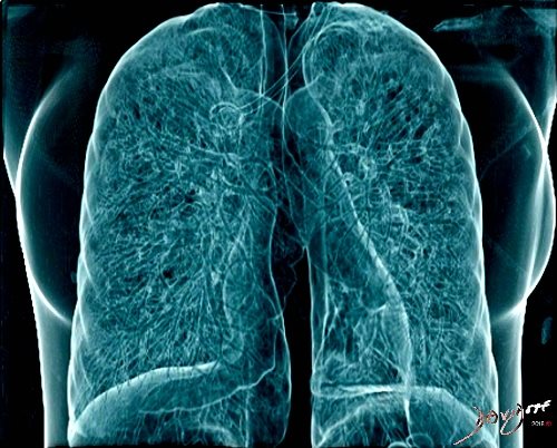
Ashley Davidoff
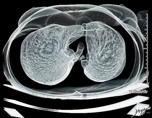
Metastatic urothelial carcinoma to the lungs
Ashley Davidoff MD
TheCommonVein.net
CT Scan
Art of Radiology
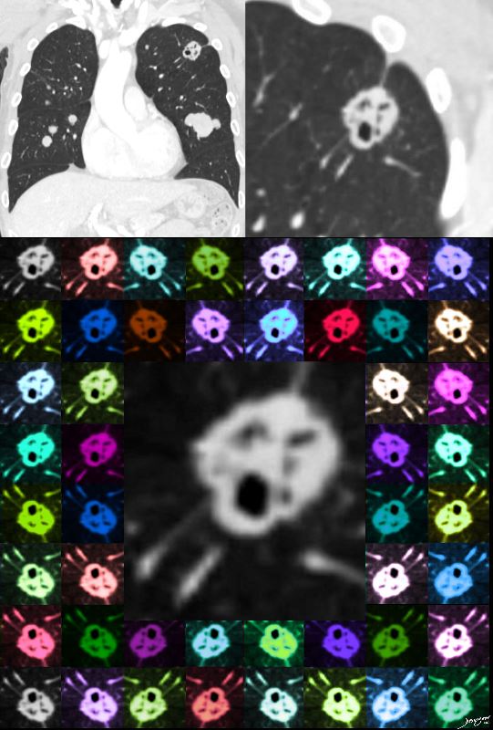
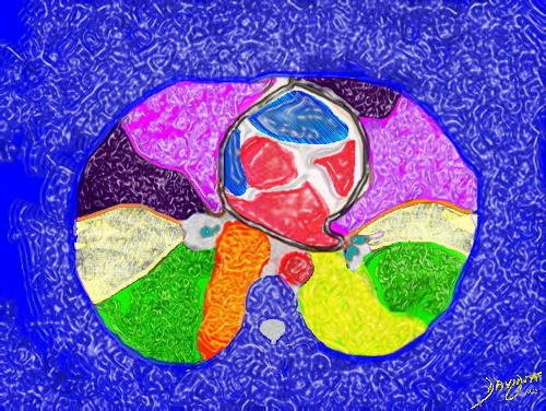
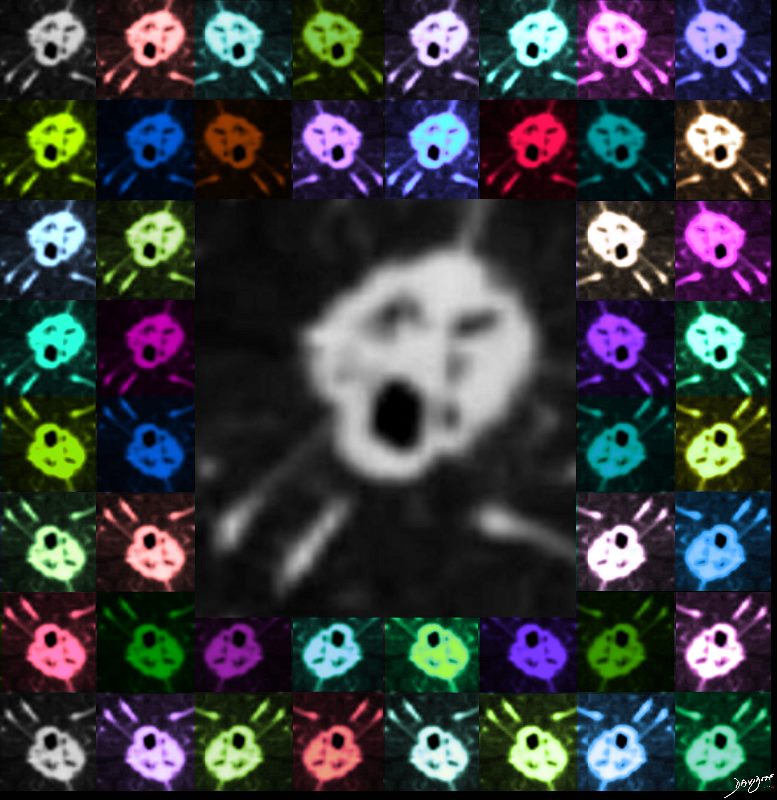
The Scream and Cry of Cavitating Metastasis
Metastatitic urothelial carcinoma
Ashley Davidoff MD
TheCommonVein.net
CT Scan
Art of Radiology

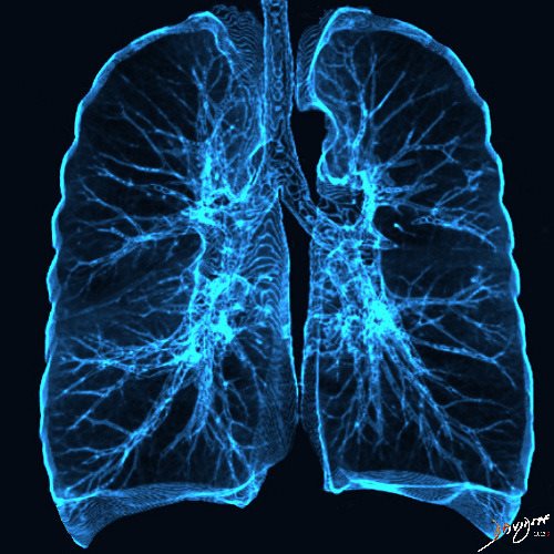
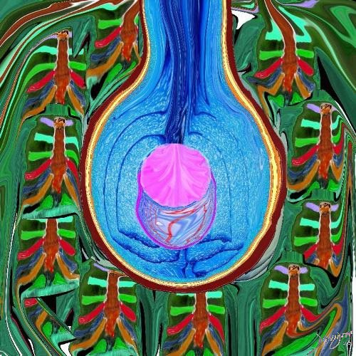
Airways turned upside down and created a tree of Gingko shaped leaves
Ashley Davidoff MD
TheCommonVein.net
CT scan
21 x 21
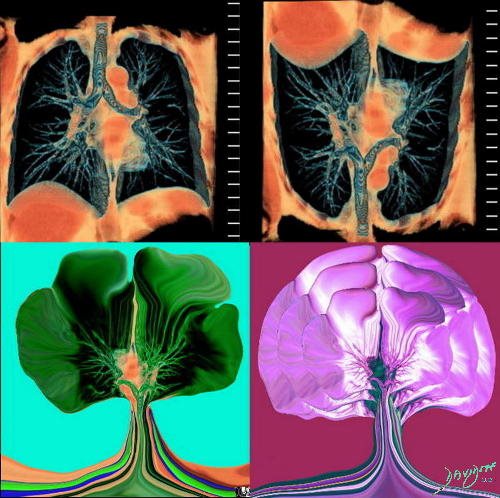
Airways turned upside down and created a tree of Gingko shaped leaves
Ashley Davidoff MD
TheCommonVein.net CT scan
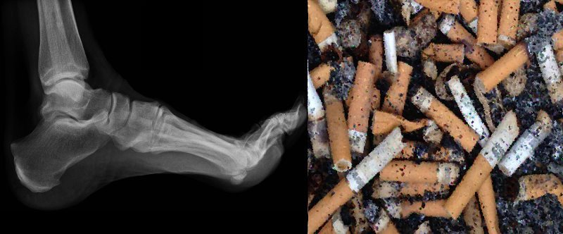
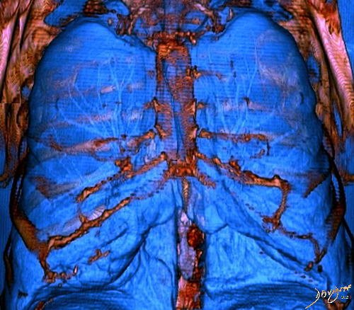
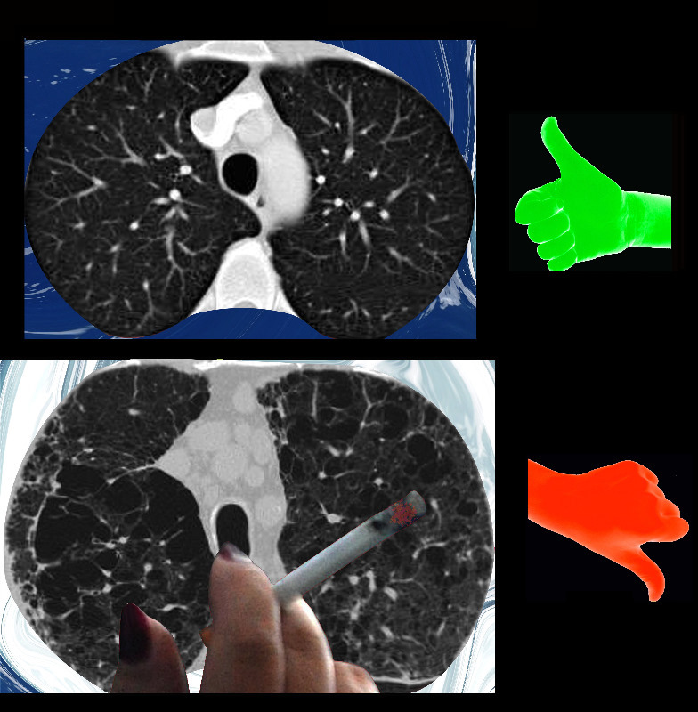
Kick the Habit in the Butt
Ashley Davidoff MD
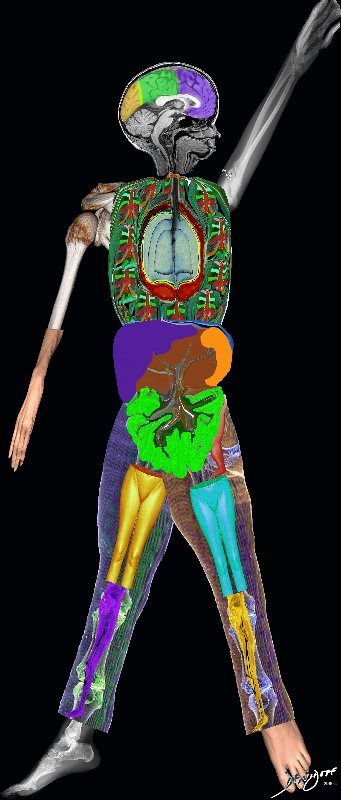
Ashley Davidoff
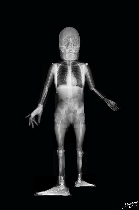
Ashley Davidoff
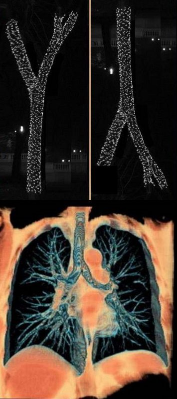
The classical branching pattern of many trees
Ashley Davidoff MD
TheCommonVein.net

Ashley Davidoff MD
TheCommonVein.net
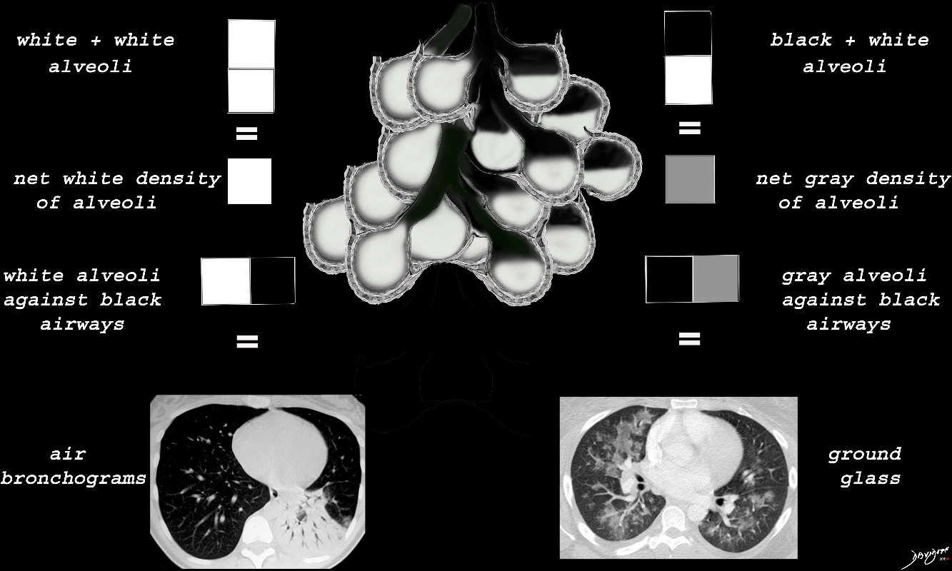
When the alveoli are fully filled with fluid, tumor, or pus for example, the overall net density will be white, and when adjacent to air filled airways, air bronchograms are visible (left side of image)
When the alveoli are only partially filled, the density of the fluid added to the density of the air results in an overall gray density, and when positioned next to air filled bronchi, there is insufficient contrast to create an air bronchogram and sufficient to enable visualization of the blood vessels. This is called ground glass opacification
Ashley Davidoff
TheCommonVein.net
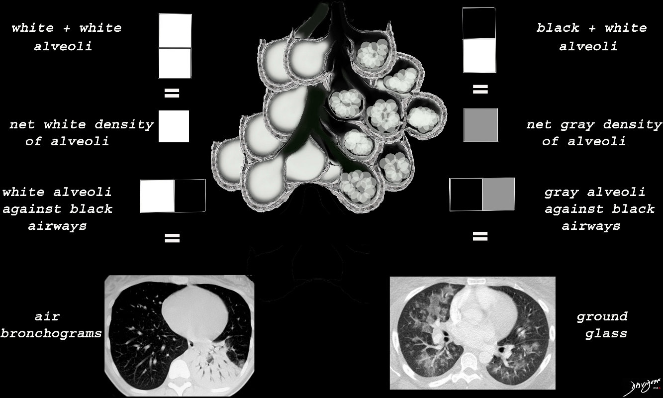
When there are extensive ceelular accumulations in the alveoli, such as adenocarcinoma with lepidic growth, Langerhans cells or other macrophages, the overall net density of the region of involvement will be gray, and when adjacent to the black air filled airways, a ground glass appearance will be apparent
Ashley Davidoff
TheCommonVein.net
ssb = subsegmental bronchiole
tb = terminal bronchiole
rb = respiratory bronchiole
as = alveolar duct
as = alvelar sac
is = anteralveolar septum
lungs-00682-lo res
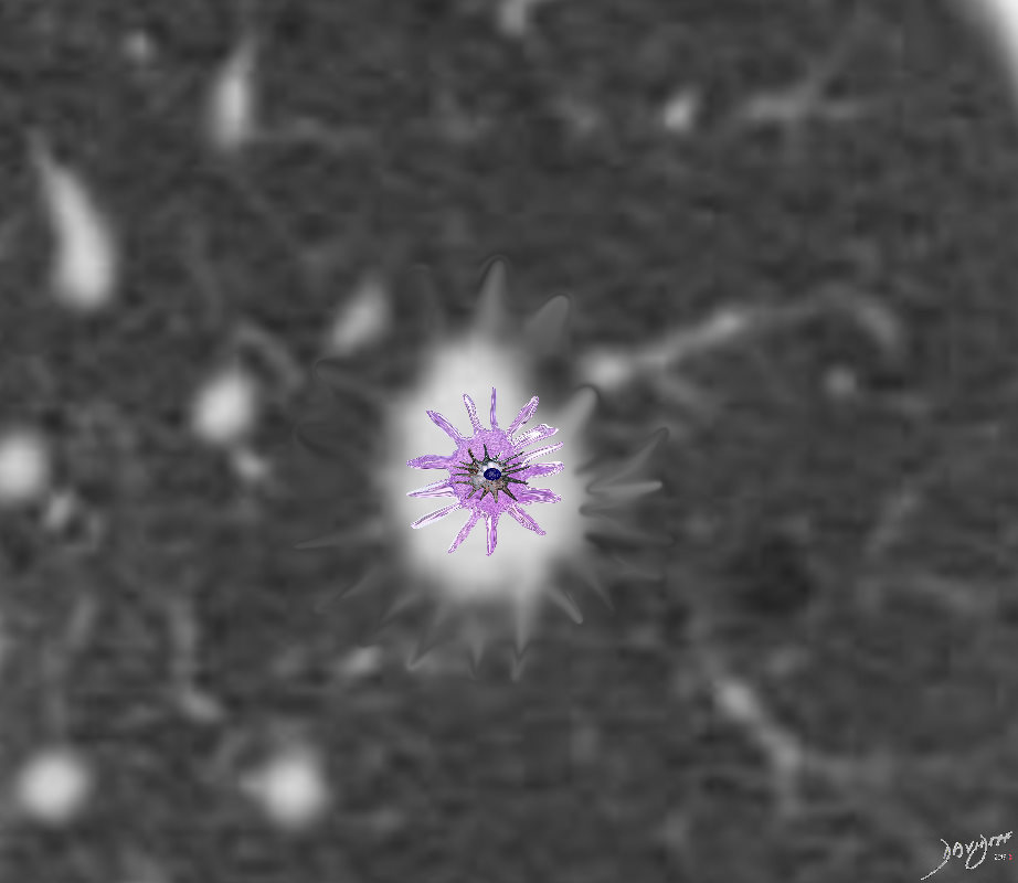
Langerhans Cell is a dendritic white cell with a wavy nucleus that creates granulomas and infiltrates the interstitium. It thus causes spiculated nodules that appear as spiculated nodules on CT
Ashley Davidoff
TheCommon Vein.net
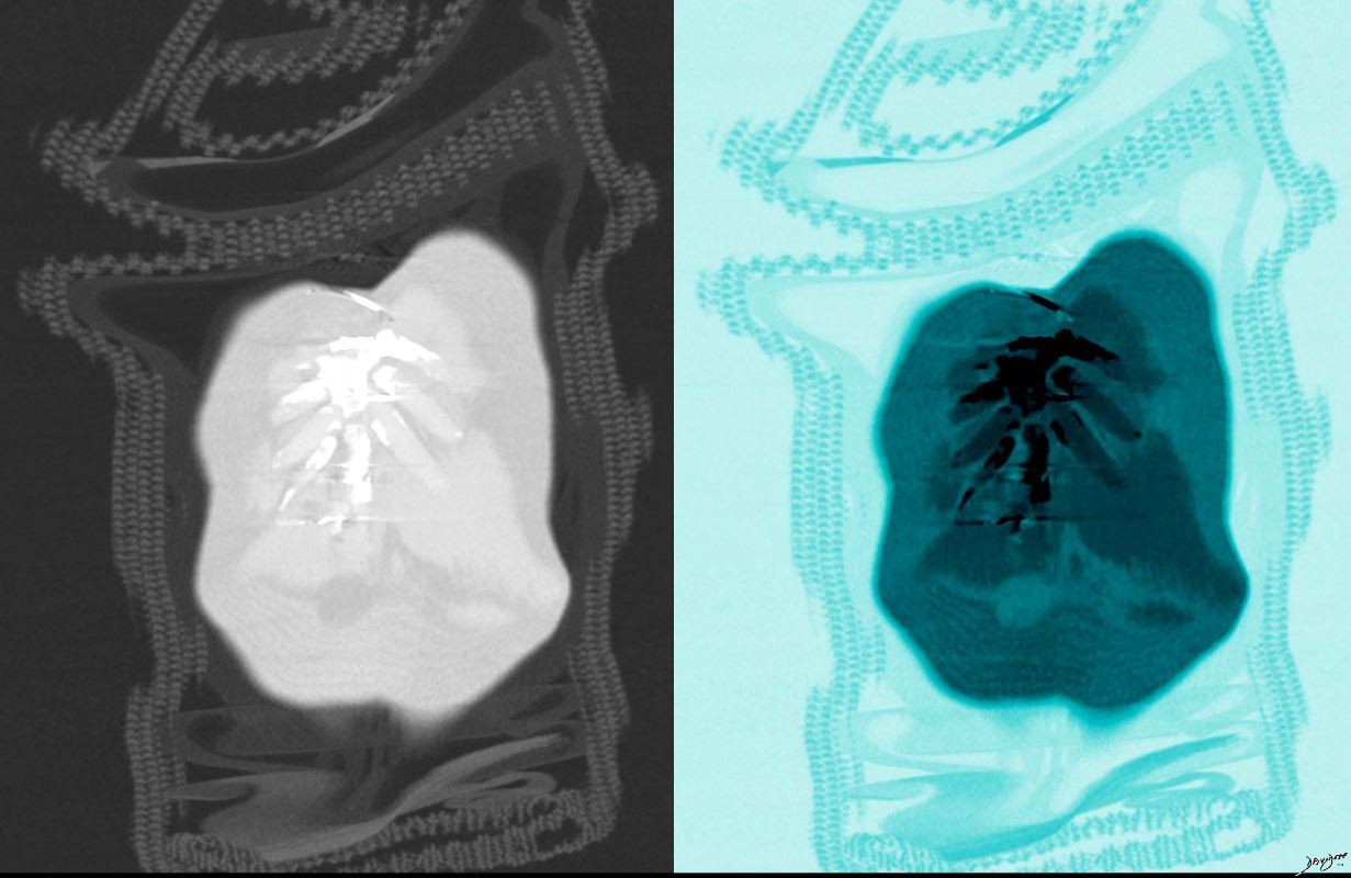
Langerhans Cell is a dendritic white cell with a wavy nucleus that creates granulomas and infiltrates the interstitium. It thus causes spiculated nodules that appear as spiculated nodules on CT
Ashley Davidoff
TheCommon Vein.net
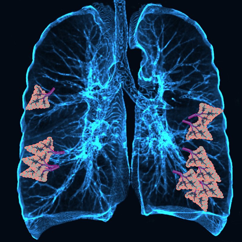
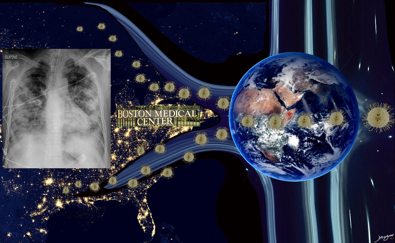
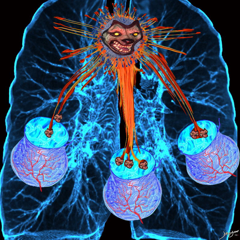
The infection starts in small basal segments
Ashley Davidoff
TheCommonVein.net
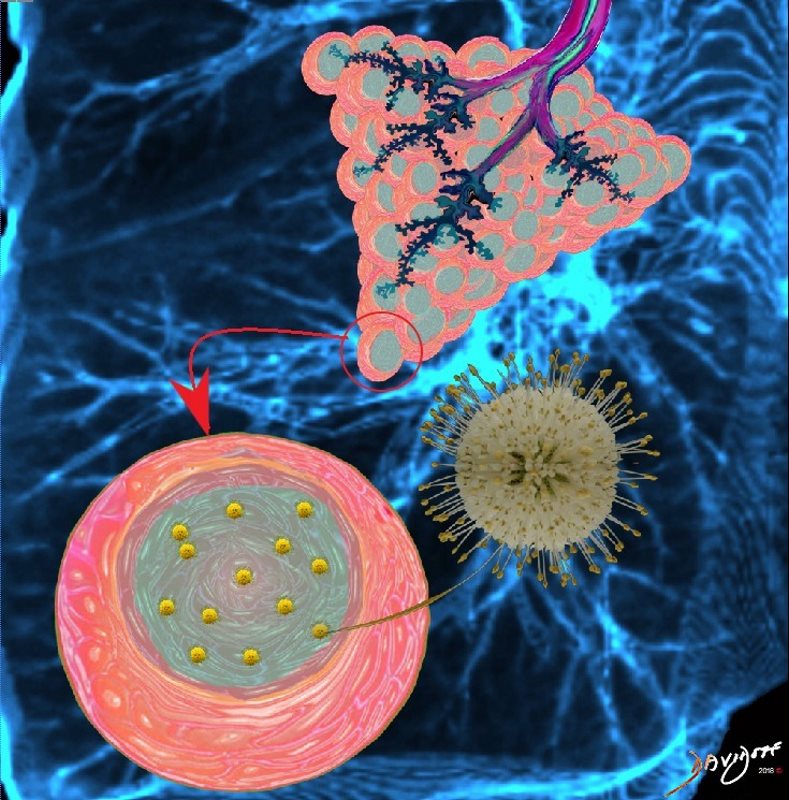
The virus replicates and invades more cells of the alveoli
As COVID-19 causes inflammation of the the lungs, infected fluid fills the lungs thus disrupting gas exchange.
Ashley Davidoff
TheCommonVein.net

3000-4,000 acini unite to form the acinus. The acini are formed by the respiratory bronchioles and the alveolar ducts, and the alveoli
Ashley Davidoff MD
TheCommonVein.net
Ashley Davidoff MD
TheCommonVein.net
Ashley Davidoff
TheCommonVein.net
